- Physical Examination
- Surgical Examination
- Ophthalmology
- Clinical Skills
- Orthopedics
- Surgery Videos
- Laparoscopy
- Pediatrics
- Funny Videos
- Cardiothoracic Surgery
- Nursing Videos
- Plastic Surgery
- Otorhinolaryngology
- Histology and Histopathology
- Neurosurgery
- Dermatology
- Pediatric Surgery
- Urology
- Dentistry
- Oncology and Cancers
- Anatomy Videos
- Health and Fitness
- Radiology
- Anaesthesia
- Physical Therapy
- Pharmacology
- Interventional Radiology
- Cardiology
- Endocrinology
- Gynecology
- Emergency Medicine
- Psychiatry and Psychology
- Childbirth Videos
- General Medical Videos
- Nephrology
- Physiology
- Diet and Food Health
- Diabetes Mellitus
- Neurology
- Women Health
- Osteoporosis
- Gastroenterology
- Pulmonology
- Hematology
- Rheumatology
- Toxicology
- Nuclear Medicine
- Infectious Diseases
- Vascular Disease
- Reproductive Health
- Burns and Wound Healing
- Kidney Zone Podcast
- Other
Talus
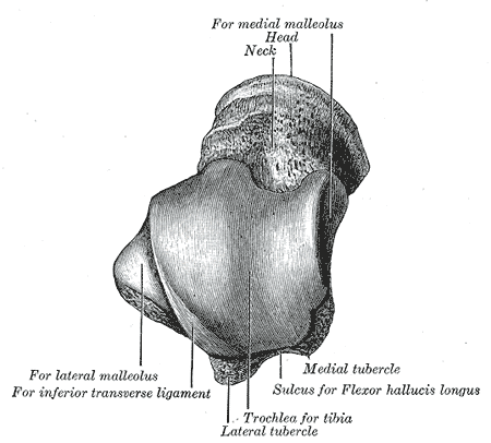
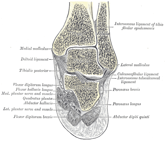
Gray's Anatomy (1918) plate, public domain
The talus is the keystone bone of the ankle: it sits between the leg bones above and the heel bone below, transmitting the entire weight of the body into the foot. Uniquely, no muscles attach to it — it is moved entirely by the bones and ligaments around it. Its domed top fits the ankle mortise formed by tibia and fibula.
Key facts
- The talus transfers all body weight from leg to foot.
- No muscle or tendon attaches to the talus.
- Most of its surface is covered in joint cartilage.
- Its blood supply enters mainly through its neck — and is easily disrupted by fractures.
- It forms the ankle joint above and the subtalar joint below.
What can go wrong
Talus neck fractures, from forced upward bending of the foot, can interrupt the bone's blood supply and lead to death of part of the bone (avascular necrosis) — which is why they are treated urgently. The talus's dome can also suffer small cartilage injuries after ankle sprains, causing deep ankle pain.
Related procedures & tests
Related symptoms
Frequently asked questions
How does the talus move with no muscles attached?
It is a passive intermediary: the leg bones drive it from above and the foot bones from below, and its shape and ligaments determine the motion. It is sometimes compared to a ball bearing in the ankle's machinery.
Why are talus fractures treated so carefully?
Because its blood vessels run backwards into the bone through the neck, a neck fracture can cut off the supply to the body of the talus, risking collapse of the ankle joint surface.
Educational information only. This atlas explains normal anatomy and describes conditions in general terms; it is not medical advice and does not replace assessment by a qualified clinician. If you have symptoms that worry you, seek medical care.