- Physical Examination
- Surgical Examination
- Ophthalmology
- Clinical Skills
- Orthopedics
- Surgery Videos
- Laparoscopy
- Pediatrics
- Funny Videos
- Cardiothoracic Surgery
- Nursing Videos
- Plastic Surgery
- Otorhinolaryngology
- Histology and Histopathology
- Neurosurgery
- Dermatology
- Pediatric Surgery
- Urology
- Dentistry
- Oncology and Cancers
- Anatomy Videos
- Health and Fitness
- Radiology
- Anaesthesia
- Physical Therapy
- Pharmacology
- Interventional Radiology
- Cardiology
- Endocrinology
- Gynecology
- Emergency Medicine
- Psychiatry and Psychology
- Childbirth Videos
- General Medical Videos
- Nephrology
- Physiology
- Diet and Food Health
- Diabetes Mellitus
- Neurology
- Women Health
- Osteoporosis
- Gastroenterology
- Pulmonology
- Hematology
- Rheumatology
- Toxicology
- Nuclear Medicine
- Infectious Diseases
- Vascular Disease
- Reproductive Health
- Burns and Wound Healing
- Kidney Zone Podcast
- Other
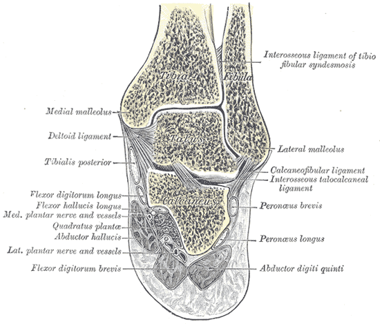
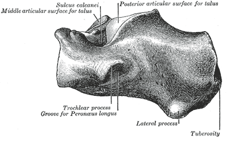
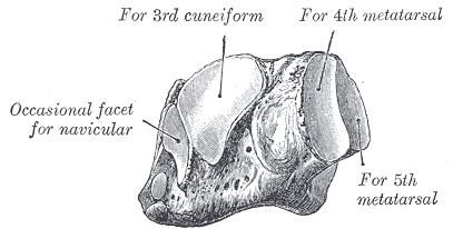
Tarsals
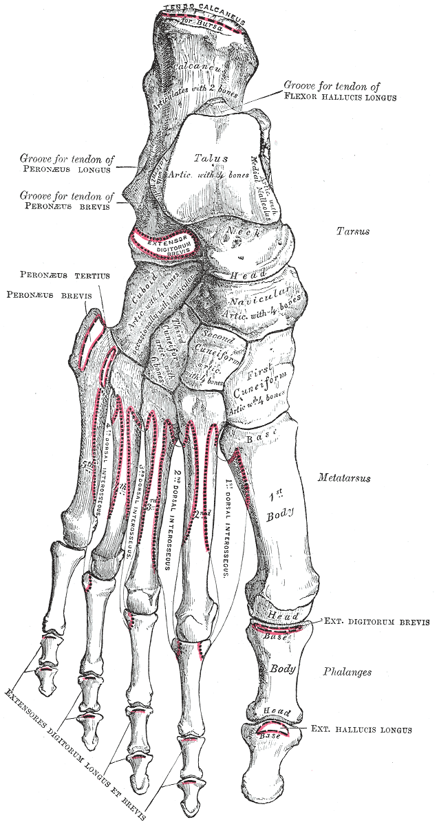
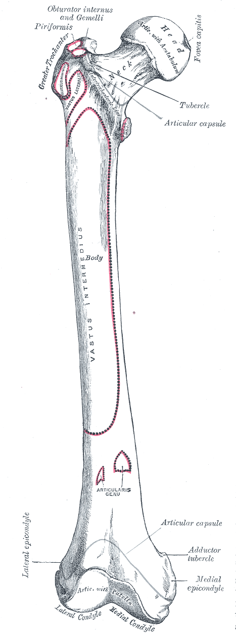
Gray's Anatomy (1918) plate, public domain
The tarsals are the seven bones of the ankle and hindfoot: the talus and calcaneus at the back, and the navicular, cuboid and three cuneiforms in front of them. Locked together by strong ligaments into the foot's arches, they transmit body weight from the leg to the ground and adapt the foot to uneven surfaces.
Key facts
- There are seven tarsals: talus, calcaneus, navicular, cuboid and three cuneiforms.
- Only the talus meets the leg bones at the ankle joint.
- They form the back half of the foot's skeleton.
- Their arrangement creates the foot's medial and lateral arches.
- The calcaneus is the largest tarsal — the heel bone.
What can go wrong
The calcaneus fractures in falls from height; the talus can fracture in forced ankle movements and has a precarious blood supply. The midfoot tarsals and their ligaments can be injured in twisting falls (Lisfranc injuries), which are important not to miss because the arch's stability depends on them.
Related procedures & tests
Related symptoms
Related muscles
Frequently asked questions
How do the tarsals differ from the carpals?
They are larger and more solidly built, arranged for weight-bearing rather than dexterity — the foot's tarsals form arches that carry the whole body, while the wrist's carpals prioritise movement.
What is the midfoot?
The navicular, cuboid and cuneiforms with the bases of the metatarsals — the keystone region of the arch. Its main joint line is called the Lisfranc joint after the surgeon who described injuries there.
Educational information only. This atlas explains normal anatomy and describes conditions in general terms; it is not medical advice and does not replace assessment by a qualified clinician. If you have symptoms that worry you, seek medical care.