- Physical Examination
- Surgical Examination
- Ophthalmology
- Clinical Skills
- Orthopedics
- Surgery Videos
- Laparoscopy
- Pediatrics
- Funny Videos
- Cardiothoracic Surgery
- Nursing Videos
- Plastic Surgery
- Otorhinolaryngology
- Histology and Histopathology
- Neurosurgery
- Dermatology
- Pediatric Surgery
- Urology
- Dentistry
- Oncology and Cancers
- Anatomy Videos
- Health and Fitness
- Radiology
- Anaesthesia
- Physical Therapy
- Pharmacology
- Interventional Radiology
- Cardiology
- Endocrinology
- Gynecology
- Emergency Medicine
- Psychiatry and Psychology
- Childbirth Videos
- General Medical Videos
- Nephrology
- Physiology
- Diet and Food Health
- Diabetes Mellitus
- Neurology
- Women Health
- Osteoporosis
- Gastroenterology
- Pulmonology
- Hematology
- Rheumatology
- Toxicology
- Nuclear Medicine
- Infectious Diseases
- Vascular Disease
- Reproductive Health
- Burns and Wound Healing
- Kidney Zone Podcast
- Other
Temporal Bone
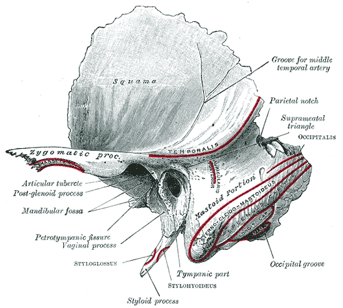
Gray's Anatomy (1918) plate, public domain
The temporal bones sit at the sides and base of the skull, level with the ears. Each houses the ear canal, the middle and inner ear, and the mastoid process — the bump you can feel behind your ear. The temporal bone also forms the socket of the jaw joint and gives attachment to neck muscles.
Key facts
- Each temporal bone surrounds and protects the structures of the ear.
- The mastoid process, behind the ear, anchors strong neck muscles such as the sternocleidomastoid.
- Its lower surface forms the socket (mandibular fossa) for the jaw joint.
- The styloid process, a slender spike below the ear, anchors tongue and throat muscles.
- The facial nerve passes through a canal in this bone on its way to the face.
What can go wrong
Temporal bone fractures can follow side-of-head injuries and may affect hearing or the facial nerve. The air cells inside the mastoid can become infected from a middle-ear infection (mastoiditis), causing pain and swelling behind the ear. This bone is also the site of the jaw joint, which can become painful or click.
Related procedures & tests
Related symptoms
Related muscles
Frequently asked questions
What is the bump behind the ear?
That is the mastoid process of the temporal bone. It is full of small air spaces connected to the middle ear, and it serves as an anchor for muscles that turn and tilt the head.
Why do doctors care about the jaw joint and the temporal bone?
Because the socket of the jaw joint (the temporomandibular joint) is part of the temporal bone, just in front of the ear — which is why jaw-joint pain is often felt as earache.
Educational information only. This atlas explains normal anatomy and describes conditions in general terms; it is not medical advice and does not replace assessment by a qualified clinician. If you have symptoms that worry you, seek medical care.