- Physical Examination
- Surgical Examination
- Ophthalmology
- Clinical Skills
- Orthopedics
- Surgery Videos
- Laparoscopy
- Pediatrics
- Funny Videos
- Cardiothoracic Surgery
- Nursing Videos
- Plastic Surgery
- Otorhinolaryngology
- Histology and Histopathology
- Neurosurgery
- Dermatology
- Pediatric Surgery
- Urology
- Dentistry
- Oncology and Cancers
- Anatomy Videos
- Health and Fitness
- Radiology
- Anaesthesia
- Physical Therapy
- Pharmacology
- Interventional Radiology
- Cardiology
- Endocrinology
- Gynecology
- Emergency Medicine
- Psychiatry and Psychology
- Childbirth Videos
- General Medical Videos
- Nephrology
- Physiology
- Diet and Food Health
- Diabetes Mellitus
- Neurology
- Women Health
- Osteoporosis
- Gastroenterology
- Pulmonology
- Hematology
- Rheumatology
- Toxicology
- Nuclear Medicine
- Infectious Diseases
- Vascular Disease
- Reproductive Health
- Burns and Wound Healing
- Kidney Zone Podcast
- Other
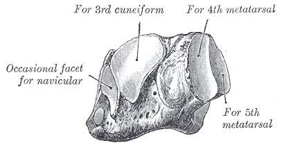
Third Cuneiform
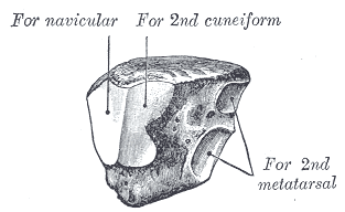
Gray's Anatomy (1918) plate, public domain
The third (lateral) cuneiform is the wedge between the second cuneiform and the cuboid, supporting the third metatarsal. With its neighbours it forms the midfoot's transverse arch and the front wall of the joints with the navicular behind. A small muscle of the big toe (flexor hallucis brevis) takes some origin from it.
Key facts
- It supports the middle (third) ray of the foot.
- It sits between the intermediate cuneiform and the cuboid.
- It completes the cuneiform row of the midfoot.
- It is part of the Lisfranc joint complex.
- The transverse arch passes through it.
What can go wrong
Like the other cuneiforms, the third is injured mainly as part of midfoot (Lisfranc) injury patterns rather than alone. Persistent pain in the top of the midfoot after a twisting injury is the presentation that prompts imaging of this whole region.
Related procedures & tests
Related symptoms
Related muscles
Frequently asked questions
Do the cuneiforms move?
Very slightly. The midfoot joints allow only a few degrees of glide — enough to let the arch flex and adapt, but the design priority is stability for weight transfer.
Why are midfoot injuries easy to miss?
Swelling can be modest and initial X-rays without weight-bearing can look near-normal. The clues are bruising under the arch and pain at the metatarsal bases, which is why loaded X-rays or CT are used when suspicion persists.
Educational information only. This atlas explains normal anatomy and describes conditions in general terms; it is not medical advice and does not replace assessment by a qualified clinician. If you have symptoms that worry you, seek medical care.