
- Physical Examination
- Surgical Examination
- Ophthalmology
- Clinical Skills
- Orthopedics
- Surgery Videos
- Laparoscopy
- Pediatrics
- Funny Videos
- Cardiothoracic Surgery
- Nursing Videos
- Plastic Surgery
- Otorhinolaryngology
- Histology and Histopathology
- Neurosurgery
- Dermatology
- Pediatric Surgery
- Urology
- Dentistry
- Oncology and Cancers
- Anatomy Videos
- Health and Fitness
- Radiology
- Anaesthesia
- Physical Therapy
- Pharmacology
- Interventional Radiology
- Cardiology
- Endocrinology
- Gynecology
- Emergency Medicine
- Psychiatry and Psychology
- Childbirth Videos
- General Medical Videos
- Nephrology
- Physiology
- Diet and Food Health
- Diabetes Mellitus
- Neurology
- Women Health
- Osteoporosis
- Gastroenterology
- Pulmonology
- Hematology
- Rheumatology
- Toxicology
- Nuclear Medicine
- Infectious Diseases
- Vascular Disease
- Reproductive Health
- Burns and Wound Healing
- Kidney Zone Podcast
- Other
Angiography
Angiography is imaging of the blood vessels, used to find narrowings, blockages, and aneurysms, and often to treat them in the same session. This page explains the catheter, CT, and MR versions and when each is chosen, in plain language with videos.
✓ Medically reviewed by Dr. Mohamed Ibrahim, MD, PhD — physician-scientist · Last reviewed: November 2026
Watch: Angiography videos
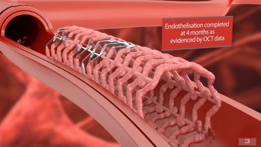 Heart Stent and Angioplasty - 3D Medical Video Animation11,892 views
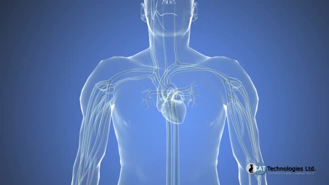 Cardiac catheterizatio5,743 views
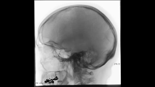 Interventional Stroke5,387 views
 Bilateral renal arterial stenosis4,015 views
Guide
What it is
In catheter angiography you lie on an X-ray table while a doctor numbs the skin — most often at the wrist or the top of the leg — and threads a fine tube (catheter) through an artery to the area being studied. Contrast dye is injected through the catheter while X-ray pictures are taken in rapid sequence (fluoroscopy), so the vessels light up as the dye flows through them. You may feel a warm flush when the dye goes in. The procedure typically takes between half an hour and an hour for diagnosis alone, longer if treatment is added.
What it shows
Angiography maps arteries and veins in fine detail: narrowed or blocked coronary arteries in heart disease, leg artery disease, aneurysms, bleeding sites, and the vessels supplying tumours or organs before surgery. The catheter version is also the platform for treatment — balloons and stents to open arteries, coils or particles to block bleeding vessels, and clot retrieval in stroke. CT and MR angiography cannot treat, but they are the usual first step for mapping, for example checking the brain's arteries or the aorta, because they are quick scans rather than procedures.
How to prepare
For a catheter angiogram you will usually fast for a few hours beforehand and have blood tests, including kidney function, checked in advance. Tell the team about previous contrast reactions, kidney problems, diabetes medication, and blood thinners — some need pausing around the procedure, and you will be told exactly which. The entry site (usually the groin or wrist) is numbed, and sedation is often given so you are relaxed but awake. Afterwards you lie flat for several hours while the puncture seals, and most people go home the same day.
What the report words mean
Angiography reports describe each vessel in turn. 'Stenosis' is a narrowing, usually given as a percentage — higher percentages mean a tighter narrowing and are used, with your symptoms, to decide on treatment. 'Occlusion' is a complete blockage. 'Collateral' vessels are natural bypass channels the body has grown around a blockage. 'Dissection' is a tear in the vessel wall lining. 'Aneurysm' is a ballooned, weakened segment. Reports end with a plan: medical treatment, angioplasty or stenting, surgery, or surveillance scans — the ordering clinician explains which applies to you.
Common questions
What is the difference between angiography and angioplasty?
A diagnostic angiogram only takes pictures. Angioplasty goes further: a balloon, and usually a stent, is used through the same catheter to open a narrowed artery during the same procedure. Many angiograms are planned with the option to treat straight away if a suitable narrowing is found.
Why a catheter angiogram instead of a CT or MR angiogram?
CT and MR angiography are scans — quick, non-invasive, and good for mapping vessels. Catheter angiography is a procedure: it gives the sharpest detail, shows flow in real time, and lets the doctor treat a narrowing in the same session. It is chosen when the scans suggest treatment is likely, or when scan pictures are not clear enough.
What is recovery like?
Afterwards you rest flat for a few hours while the puncture site seals, with checks on the site and the pulse beyond it. Most people go home the same day or the next, should avoid heavy lifting for a few days, and are told to seek help promptly for bleeding, swelling, or a cold or painful limb.
Can I react to the dye?
Yes — a small number of people have an allergic-type reaction to the dye, from itching and a rash to, rarely, a severe reaction. The team asks about previous reactions in advance, can pre-treat or choose alternatives, and is equipped to treat reactions immediately if they happen.
More in Angiography & Interventional
Related
Sources
- Angiography (NHS)
- Catheter Angiography (RadiologyInfo.org (RSNA/ACR))
- Coronary angiogram (MedlinePlus Medical Encyclopedia)
This guide is general information, not medical advice, and does not replace advice from your own clinician. How a scan is done, and whether contrast is used, depends on your own medical history — always follow the instructions of the team performing your scan, and tell them about kidney problems, allergies, implants, and the medicines you take. Angiography through a catheter is a procedure with small risks of bleeding, vessel injury, and contrast reactions — your team will explain the balance of risks and benefits for your case.