- Physical Examination
- Surgical Examination
- Ophthalmology
- Clinical Skills
- Orthopedics
- Surgery Videos
- Laparoscopy
- Pediatrics
- Funny Videos
- Cardiothoracic Surgery
- Nursing Videos
- Plastic Surgery
- Otorhinolaryngology
- Histology and Histopathology
- Neurosurgery
- Dermatology
- Pediatric Surgery
- Urology
- Dentistry
- Oncology and Cancers
- Anatomy Videos
- Health and Fitness
- Radiology
- Anaesthesia
- Physical Therapy
- Pharmacology
- Interventional Radiology
- Cardiology
- Endocrinology
- Gynecology
- Emergency Medicine
- Psychiatry and Psychology
- Childbirth Videos
- General Medical Videos
- Nephrology
- Physiology
- Diet and Food Health
- Diabetes Mellitus
- Neurology
- Women Health
- Osteoporosis
- Gastroenterology
- Pulmonology
- Hematology
- Rheumatology
- Toxicology
- Nuclear Medicine
- Infectious Diseases
- Vascular Disease
- Reproductive Health
- Burns and Wound Healing
- Kidney Zone Podcast
- Other
Intervertebral Discs
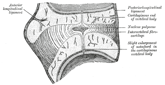
Gray's Anatomy (1918) plate, public domain
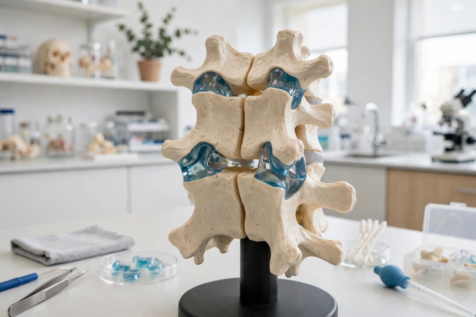
Intervertebral discs are the tough cushions between the vertebrae — 23 of them from neck to lower back. Each has a firm outer ring of fibrous cartilage (annulus fibrosus) around a soft, jelly-like centre (nucleus pulposus). Together they absorb shock, hold the vertebrae together and give the spine about a quarter of its length.
Key facts
- Discs contribute about 25% of the spine's total length.
- The outer annulus is made of concentric layers of tough fibres.
- The nucleus is mostly water, which is why discs lose a little height over the day.
- Discs have almost no blood supply after childhood and heal slowly.
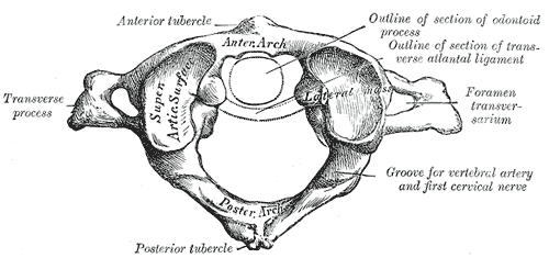
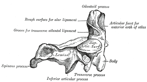
- There is no disc between the skull and the first two neck vertebrae.
What can go wrong
With age the nucleus dries out and the annulus can develop tears, so discs narrow and bulge. A prolapsed ('slipped') disc can press on a spinal nerve, causing pain, pins and needles or weakness down an arm or leg. Most disc problems settle without surgery; new bladder or bowel disturbance with back pain is an emergency sign.
Related procedures & tests
Related symptoms
Frequently asked questions
What does 'slipped disc' actually mean?
Discs do not slip — the term describes a bulge or herniation, where the soft centre pushes through a tear in the outer ring and can press on a nearby nerve.
Why am I taller in the morning?
Overnight, unloaded discs reabsorb fluid and swell slightly; during the day gravity squeezes some water out again. The difference can be up to about 2 centimetres across the whole spine.
Educational information only. This atlas explains normal anatomy and describes conditions in general terms; it is not medical advice and does not replace assessment by a qualified clinician. If you have symptoms that worry you, seek medical care.