- Physical Examination
- Surgical Examination
- Ophthalmology
- Clinical Skills
- Orthopedics
- Surgery Videos
- Laparoscopy
- Pediatrics
- Funny Videos
- Cardiothoracic Surgery
- Nursing Videos
- Plastic Surgery
- Otorhinolaryngology
- Histology and Histopathology
- Neurosurgery
- Dermatology
- Pediatric Surgery
- Urology
- Dentistry
- Oncology and Cancers
- Anatomy Videos
- Health and Fitness
- Radiology
- Anaesthesia
- Physical Therapy
- Pharmacology
- Interventional Radiology
- Cardiology
- Endocrinology
- Gynecology
- Emergency Medicine
- Psychiatry and Psychology
- Childbirth Videos
- General Medical Videos
- Nephrology
- Physiology
- Diet and Food Health
- Diabetes Mellitus
- Neurology
- Women Health
- Osteoporosis
- Gastroenterology
- Pulmonology
- Hematology
- Rheumatology
- Toxicology
- Nuclear Medicine
- Infectious Diseases
- Vascular Disease
- Reproductive Health
- Burns and Wound Healing
- Kidney Zone Podcast
- Other
Nasal Septum
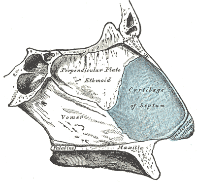
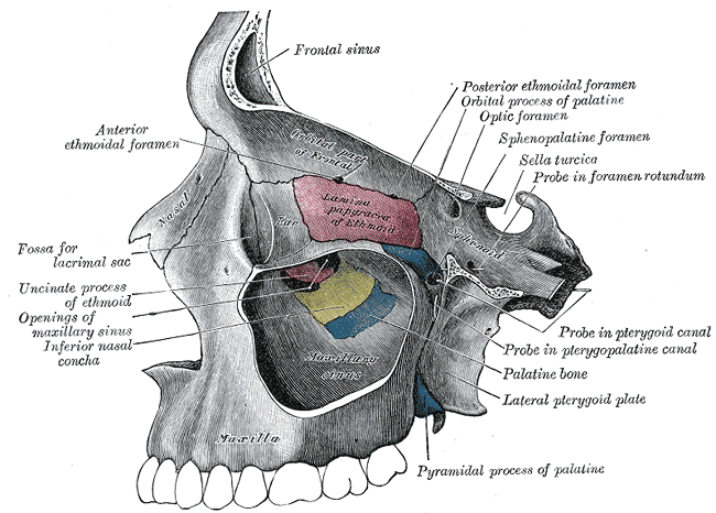
Gray's Anatomy (1918) plate, public domain
The nasal septum is the wall dividing the nose into two passages, made of cartilage at the front and thin bone (the perpendicular plate of the ethmoid and the vomer) behind, all covered by mucosal lining. Its front lower lining (Kiesselbach's area) carries a rich plexus of small blood vessels — the commonest site of nosebleeds. A perfectly straight septum is uncommon; deviations are present in most adults to some degree.
Key facts
- The septum's cartilage continues to grow through adolescence, which is why childhood deviations can evolve.
- Its rich blood supply at the front makes it the source of the great majority of nosebleeds.
- The septum supports the nose's shape — damage to it can cause the bridge to sag.
- Septal cartilage is a favourite donor material for reconstructive surgery elsewhere in the nose and ear.
What can go wrong
A significantly deviated septum — from development or injury — can block one nostril, disturb airflow and contribute to snoring, sinus symptoms and nosebleeds; corrective surgery is called septoplasty. Bleeding from its front vessels is managed with first aid and sometimes cautery. Rarely, the septum is perforated (from surgery, trauma, infection or cocaine use), causing crusting, whistling and bleeding. A blood collection under its lining after injury (septal haematoma) needs prompt drainage to protect the cartilage.
Related procedures & tests
Related symptoms
Watch: related videos
Frequently asked questions
Is a deviated septum abnormal?
Some degree of deviation is present in most people and is a normal variant; it matters only when it causes symptoms such as persistent one-sided blockage or recurrent nosebleeds.
Why do most nosebleeds come from the septum?
Four or five arteries converge in a fragile plexus on its front lower part (Kiesselbach's area), where the lining is thin and exposed to dry air and fingers.
Educational information only. This atlas explains normal anatomy and describes conditions in general terms; it is not medical advice and does not replace assessment by a qualified clinician. If you have symptoms that worry you, seek medical care.