- Physical Examination
- Surgical Examination
- Ophthalmology
- Clinical Skills
- Orthopedics
- Surgery Videos
- Laparoscopy
- Pediatrics
- Funny Videos
- Cardiothoracic Surgery
- Nursing Videos
- Plastic Surgery
- Otorhinolaryngology
- Histology and Histopathology
- Neurosurgery
- Dermatology
- Pediatric Surgery
- Urology
- Dentistry
- Oncology and Cancers
- Anatomy Videos
- Health and Fitness
- Radiology
- Anaesthesia
- Physical Therapy
- Pharmacology
- Interventional Radiology
- Cardiology
- Endocrinology
- Gynecology
- Emergency Medicine
- Psychiatry and Psychology
- Childbirth Videos
- General Medical Videos
- Nephrology
- Physiology
- Diet and Food Health
- Diabetes Mellitus
- Neurology
- Women Health
- Osteoporosis
- Gastroenterology
- Pulmonology
- Hematology
- Rheumatology
- Toxicology
- Nuclear Medicine
- Infectious Diseases
- Vascular Disease
- Reproductive Health
- Burns and Wound Healing
- Kidney Zone Podcast
- Other
Patella
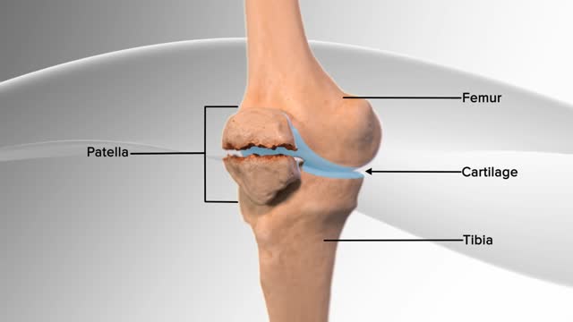
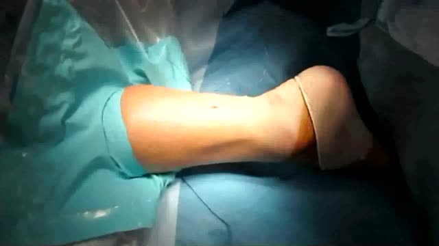
Video still from MedicalVideos.com: patella fracture repair
The patella, or kneecap, is a flat triangular sesamoid bone embedded in the tendon of the quadriceps, just in front of the knee. It glides in a groove on the femur as the knee bends and works as a pulley: by holding the tendon away from the joint, it increases the thigh muscle's leverage for straightening the knee.
Key facts
- The patella is the largest sesamoid bone in the body.
- It increases the quadriceps' leverage by roughly a third.
- Its back surface has the thickest cartilage in the body.
- It protects the front of the knee joint.
- It starts to ossify in early childhood and is fully formed by the teens.
What can go wrong
The kneecap can dislocate sideways, most often in teenagers, when a twist pushes it out of its groove. It can fracture from a direct fall onto the knee. Pain behind the kneecap on stairs and after sitting ('movie-goer's knee') commonly comes from irritation of its joint surface and the structures around it.
Related procedures & tests
Related symptoms
Related muscles
Watch: related videos
Frequently asked questions
Can you manage without a kneecap?
People do function after its removal, but the thigh muscle loses leverage, so straightening the knee against resistance is weaker — which is why surgeons preserve or repair it whenever possible.
Why does the kneecap sometimes slip out?
Its groove can be shallow, the thigh muscles unbalanced, or ligaments loose — a twisting injury then lets it jump sideways, usually to the outer side. It often pops back as the knee straightens.
Educational information only. This atlas explains normal anatomy and describes conditions in general terms; it is not medical advice and does not replace assessment by a qualified clinician. If you have symptoms that worry you, seek medical care.