- Physical Examination
- Surgical Examination
- Ophthalmology
- Clinical Skills
- Orthopedics
- Surgery Videos
- Laparoscopy
- Pediatrics
- Funny Videos
- Cardiothoracic Surgery
- Nursing Videos
- Plastic Surgery
- Otorhinolaryngology
- Histology and Histopathology
- Neurosurgery
- Dermatology
- Pediatric Surgery
- Urology
- Dentistry
- Oncology and Cancers
- Anatomy Videos
- Health and Fitness
- Radiology
- Anaesthesia
- Physical Therapy
- Pharmacology
- Interventional Radiology
- Cardiology
- Endocrinology
- Gynecology
- Emergency Medicine
- Psychiatry and Psychology
- Childbirth Videos
- General Medical Videos
- Nephrology
- Physiology
- Diet and Food Health
- Diabetes Mellitus
- Neurology
- Women Health
- Osteoporosis
- Gastroenterology
- Pulmonology
- Hematology
- Rheumatology
- Toxicology
- Nuclear Medicine
- Infectious Diseases
- Vascular Disease
- Reproductive Health
- Burns and Wound Healing
- Kidney Zone Podcast
- Other
Pericardium
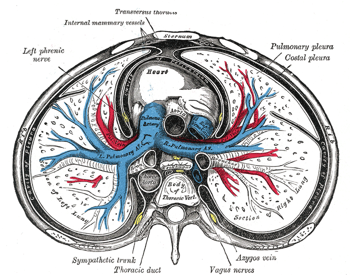
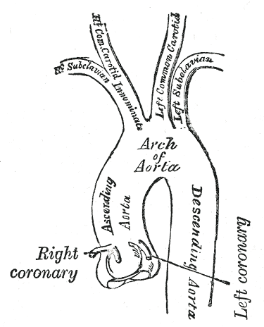
Gray's Anatomy (1918) plate, public domain
The pericardium is the double-walled sac enclosing the heart. Its tough outer fibrous layer anchors the heart in the chest; its inner serous layers coat the heart and line the sac, with a film of fluid between them so the heart beats almost friction-free. The sac limits over-expansion, keeps the heart in position and forms a barrier against infection spreading from nearby structures.
Key facts
- The pericardial space normally holds only 15-50 mL of fluid.
- The phrenic nerves run over its surface on their way to the diaphragm — a relation that matters in surgery.
- Its inner layer is continuous with the coating of the great vessels at the heart's base.
- The sac is pain-sensitive, which is why pericardial inflammation causes chest pain.
What can go wrong
Inflammation (pericarditis) causes sharp central chest pain that is characteristically worse lying flat and better sitting forward, often after a viral illness. Fluid collecting in the sac (pericardial effusion) may be silent, but if it accumulates fast it can compress the heart (tamponade) — a life-threatening emergency with breathlessness and falling blood pressure. Long-standing inflammation can scar the sac into a rigid shell (constrictive pericarditis).
Related procedures & tests
Related symptoms
Frequently asked questions
What is cardiac tamponade?
Compression of the heart by fluid under pressure in the pericardial sac, preventing it filling; it is treated as an emergency and relieved by draining the fluid.
Why does pericarditis pain change with position?
The inflamed sac rubs and stretches against surrounding structures; lying flat increases contact and pressure, while leaning forward relieves it — a classic distinguishing feature.
Educational information only. This atlas explains normal anatomy and describes conditions in general terms; it is not medical advice and does not replace assessment by a qualified clinician. If you have symptoms that worry you, seek medical care.