- Physical Examination
- Surgical Examination
- Ophthalmology
- Clinical Skills
- Orthopedics
- Surgery Videos
- Laparoscopy
- Pediatrics
- Funny Videos
- Cardiothoracic Surgery
- Nursing Videos
- Plastic Surgery
- Otorhinolaryngology
- Histology and Histopathology
- Neurosurgery
- Dermatology
- Pediatric Surgery
- Urology
- Dentistry
- Oncology and Cancers
- Anatomy Videos
- Health and Fitness
- Radiology
- Anaesthesia
- Physical Therapy
- Pharmacology
- Interventional Radiology
- Cardiology
- Endocrinology
- Gynecology
- Emergency Medicine
- Psychiatry and Psychology
- Childbirth Videos
- General Medical Videos
- Nephrology
- Physiology
- Diet and Food Health
- Diabetes Mellitus
- Neurology
- Women Health
- Osteoporosis
- Gastroenterology
- Pulmonology
- Hematology
- Rheumatology
- Toxicology
- Nuclear Medicine
- Infectious Diseases
- Vascular Disease
- Reproductive Health
- Burns and Wound Healing
- Kidney Zone Podcast
- Other
Vestibulocochlear Nerve
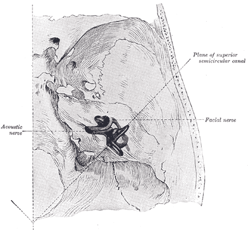
Gray's Anatomy (1918) plate, public domain
The vestibulocochlear nerve (cranial nerve VIII) carries hearing and balance information from the inner ear to the brainstem. It has two divisions: the cochlear nerve, from the spiral hearing organ, and the vestibular nerve, from the balance canals and gravity sensors. It enters the skull through the internal ear canal alongside the facial nerve.
Key facts
- It is a purely sensory nerve with two distinct divisions.
- The cochlear division carries around 30,000 fibres from each ear.
- The vestibular division reports head rotation and linear acceleration.
- It travels with the facial nerve through the internal acoustic meatus.
- It is the nerve affected by vestibular schwannoma (acoustic neuroma).
What can go wrong
A benign tumour of this nerve's sheath — vestibular schwannoma — typically causes slowly progressive one-sided hearing loss and tinnitus. Inflammation of the vestibular division (vestibular neuritis) causes sudden prolonged vertigo after a viral illness. Sudden one-sided hearing loss is treated as an emergency because early treatment gives the best chance of recovery.
Related procedures & tests
Related symptoms
Frequently asked questions
Why do hearing and balance share a nerve?
Both senses live in the same organ — the inner ear — where the cochlea and the balance apparatus sit side by side in the temporal bone, so their fibres naturally travel together.
What is an acoustic neuroma?
A usually slow-growing benign tumour on the vestibular part of this nerve (more precisely a vestibular schwannoma). Most are managed by monitoring scans; some are treated with focused radiotherapy or surgery.
Educational information only. This atlas explains normal anatomy and describes conditions in general terms; it is not medical advice and does not replace assessment by a qualified clinician. If you have symptoms that worry you, seek medical care.