
- Procedures & Tests
- Symptoms A-Z
- Symptom Checker
- Kidney Diet
- First Aid
- Anatomy Atlas
- Ask MedicalVideos
- Drug Guide
- Lab Values
- Vital Signs
- Muscles A–Z
- Clinical Signs
- Medical Abbreviations
- Medical Terminology
- Physical Examination
- Surgical Examination
- Ophthalmology
- Clinical Skills
- Orthopedics
- Surgery Videos
- Laparoscopy
- Pediatrics
- Medical Humor
- Cardiothoracic Surgery
- Nursing Videos
- Plastic Surgery
- Otorhinolaryngology
- Histology and Histopathology
- Neurosurgery
- Dermatology
- Pediatric Surgery
- Urology
- Dentistry
- Oncology and Cancers
- Anatomy Videos
- Health and Fitness
- Radiology
- Anaesthesia
- Physical Therapy
- Pharmacology
- Interventional Radiology
- Cardiology
- Endocrinology
- Gynecology
- Emergency Medicine
- Psychiatry and Psychology
- Childbirth Videos
- General Medical Videos
- Nephrology
- Physiology
- Diet and Food Health
- Diabetes Mellitus
- Neurology
- Women Health
- Osteoporosis
- Gastroenterology
- Pulmonology
- Hematology
- Rheumatology
- Toxicology
- Nuclear Medicine
- Infectious Diseases
- Vascular Disease
- Reproductive Health
- Burns and Wound Healing
- Kidney Zone Podcast
- Other

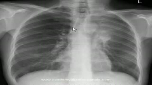
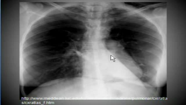
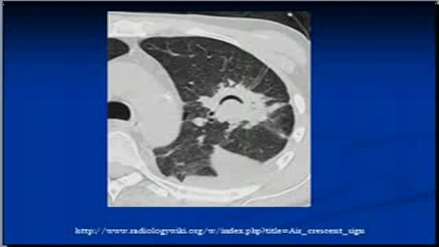






Chest x-ray interpretation, RDS

Educational only: videos on MedicalVideos.com are for general information and are not medical advice. Always consult your own doctor about your health.
The video will describe RDS in premature babies. Please see website for disclaimer
This video in 30 seconds
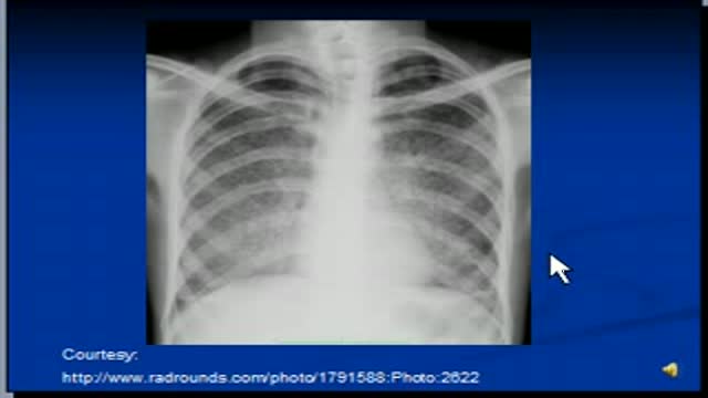
A radiology lecture examines chest X-rays of a premature baby with respiratory distress syndrome over the first days of life. The lecturer explains why preterm infants lack surfactant, then follows the films as a tension pneumothorax and pulmonary interstitial emphysema develop. Tubes visible on the films, including a nasogastric tube and an intercostal drain, are pointed out along the way.
Key points
- The lecturer explains surfactant deficiency in premature infants
- The day-one film is described as showing haziness suggesting respiratory distress syndrome
- The day-two film shows a right-sided tension pneumothorax with a shifted mediastinum
- The day-three film shows an intercostal drain and an endotracheal tube in place
- A bubbly appearance in the left lung is identified as pulmonary interstitial emphysema
Chapters
- 0:00 — Respiratory distress syndrome and surfactant
- 1:54 — Day-one chest X-ray findings
- 2:22 — Day two: tension pneumothorax
- 3:03 — Day three: intercostal drain placed
- 4:45 — Pulmonary interstitial emphysema explained
Video Transcript
A radiology lecture on chest X-rays in a premature baby with respiratory distress syndrome (RDS). The lecturer explains why preterm infants lack surfactant, then walks through the day-one, day-two and day-three films as the RDS is complicated by a tension pneumothorax and pulmonary interstitial emphysema, pointing out the nasogastric tube and the intercostal drain along the way. It is one of the DTW Academy lecture series.
Read the full transcript
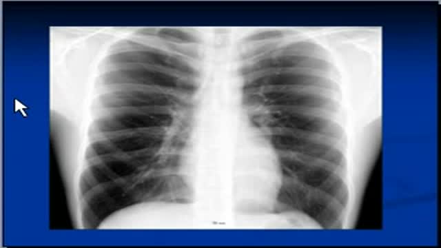
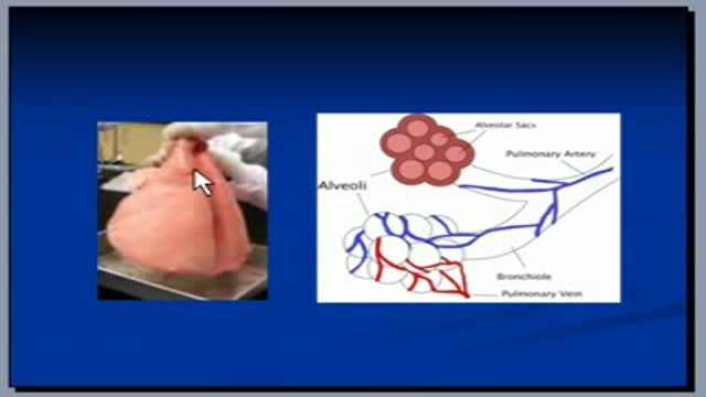
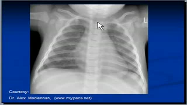
This is the next day of a premature baby born at 32 weeks of gestation, guaranteeing after 4 hours of birth x-ray taken on day 1, Query RDS or Respiratory Distress Syndrome. Respiratory Distress Syndrome in premature babies is a common problem. In mature births, a chemical called surfactant keeps alveoli open so that they easily fill with air and work efficiently. Surfactant is made by type 2 nemocytes cells of lungs and lungs do not start making surfactant until later in pregnancy. So premature babies cannot keep their alveoli open as full-terms babies do.
Such babies have to work very hard to meet their oxygen requirement. The condition is known as Respiratory Distress Syndrome or RDS. The symptoms are difficulty in breathing, sinus which is a bluish color of the skin, layering of nostrils, rapid breathing which is known as stichypnea, granting sounds with breathing and chest retractions which is pulled off ribs and sternum inside during breathing. There are two major complications of RDS. One is nemothorics which means air in the plural cavity, pressure on lungs from babies' own efforts to breathe or from the artificial breathing machine which is known as ventilator
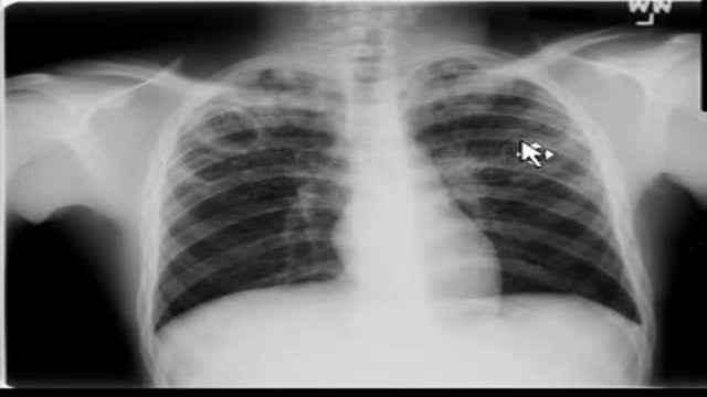
can cause lungs to rupture which causes air to leak into the plural cavity. This air causes lung to collapse. The other complication is intraventricular hemorrhage which is bleeding into the ventricles of the brain. So X-ray taken on day one does not show much to an inexperienced eye but experienced a radiologist would argue that there is a slight haziness of lungs to suggest RDS. There is an NG tube, nasogastric tube in position. The tube is inserted from nose, passes through the esophagus and goes into the stomach mainly for feeding purpose.
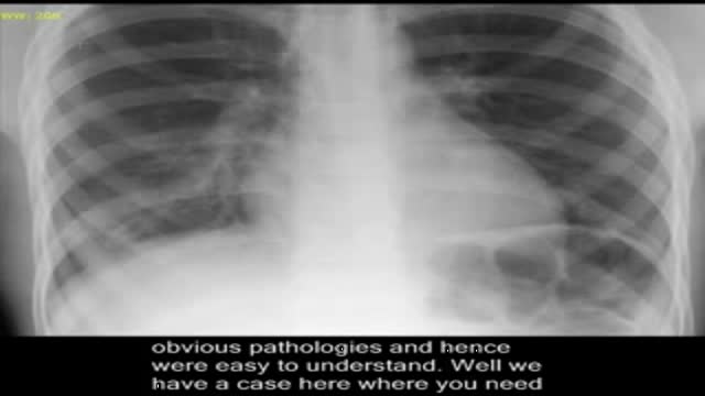
So chest X-ray on day two shows right-sided tension nemothorics in less aerated lungs indicating worsening of RDS. We call it tension nemothorics because the mediasinum is pushed towards the left-hand side and the collapsed lung is visible here. So this is free air into the plural cavity which is known as nemothorics. Another chest X-ray on day two shows needle in the plural cavity. NG tube has retracted into distal esophagus and should be advanced. Chest X-ray day three. First thing that you notice in this X-ray is this intercostal tube.
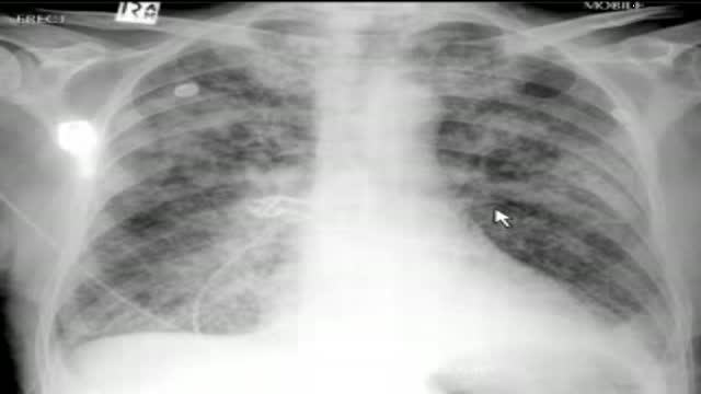
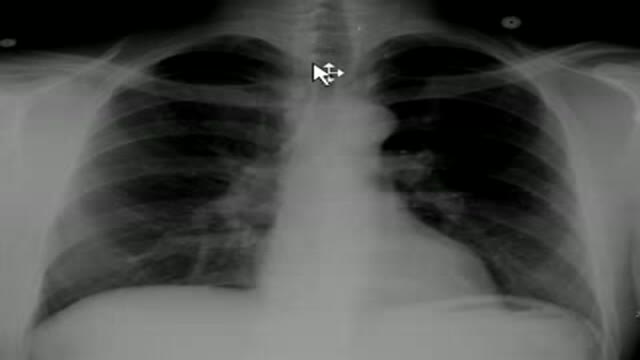
Means there is a tube that is going through the ribs between two ribs, intercostal which is normally attached to a suction on the other end to pull air from the plural cavity and to expand lung. Endotracheal tube is also visible means that patient is on artificial breathing. The tube is inserted from mouth and goes into trachea and is attached to a vent later which pushes oxygen into lungs and pulls carbon dioxide. Nezogastric tube is also in satisfactory position. So the tube has drained the nemothorics and although the lung is still partially collapsed
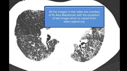
notice bubbly appearance of left lung which suggests pulmonary interstitial amperesema. Chest X-ray day five. Intercostal tube has been removed and right lung is now almost fully expanded. Satisfactory position of end of regular tube which is almost in the middle of right here. The NG tube should be at most the bubbly appearance of the left lung again suggesting pulmonary interstitial amperesema and we will discuss what is pulmonary interstitial amperesema. Pulmonary interstitial amperesema is a collection of gas outside of the normal air pathages. So air goes into the interstitial tissue and is secondary to alveolar and terminal broncholary rupture.
Pulmonary interstitial amperesema is more frequent in premature infants who require mechanical ventilation and it often occurs in conjunction with respiratory distress syndrome. The lecture is prepared for the DTW Academy of Professions.com. These are the references and X-rays are courtesy of Dr. Alex McLennan who is a radiologist in Royal Alexander Hospital, United Kingdom. Thank you very much.
Transcript auto-generated from the video’s audio for accessibility and study. Educational content only — not medical advice.
Related reading
Save videos for later
Create a free account to build your own watch-later list and playlists, and pick up where you left off on any device.
Medical Videos
Free medical and surgery videos for patients, students and professionals — trusted since 2007.
Founded by Dr. Mohamed Ibrahim, MD, PhD — physician-scientist.
Follow us on Facebook · Instagram
SORT BY-
Top Comments
-
Latest comments