
- Procedures & Tests
- Symptoms A-Z
- Symptom Checker
- Kidney Diet
- First Aid
- Anatomy Atlas
- Ask MedicalVideos
- Drug Guide
- Lab Values
- Vital Signs
- Muscles A–Z
- Clinical Signs
- Medical Abbreviations
- Medical Terminology
- Physical Examination
- Surgical Examination
- Ophthalmology
- Clinical Skills
- Orthopedics
- Surgery Videos
- Laparoscopy
- Pediatrics
- Funny Videos
- Cardiothoracic Surgery
- Nursing Videos
- Plastic Surgery
- Otorhinolaryngology
- Histology and Histopathology
- Neurosurgery
- Dermatology
- Pediatric Surgery
- Urology
- Dentistry
- Oncology and Cancers
- Anatomy Videos
- Health and Fitness
- Radiology
- Anaesthesia
- Physical Therapy
- Pharmacology
- Interventional Radiology
- Cardiology
- Endocrinology
- Gynecology
- Emergency Medicine
- Psychiatry and Psychology
- Childbirth Videos
- General Medical Videos
- Nephrology
- Physiology
- Diet and Food Health
- Diabetes Mellitus
- Neurology
- Women Health
- Osteoporosis
- Gastroenterology
- Pulmonology
- Hematology
- Rheumatology
- Toxicology
- Nuclear Medicine
- Infectious Diseases
- Vascular Disease
- Reproductive Health
- Burns and Wound Healing
- Kidney Zone Podcast
- Other
Cardiology
Cardiology videos: ECG interpretation, echocardiograms, angioplasty, stents and heart conditions explained by cardiologists. Free to watch and learn.
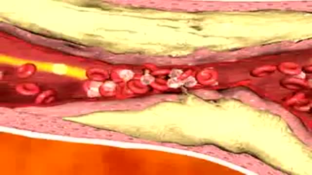

In this animation it depicts an artery with cholesterol plaque (the yellowish area) which is blocking most of the inside of this artery. Then small cells called platelets become clumped together and cause the red blood cells to form a clot. This is what causes a HEART ATTACK.
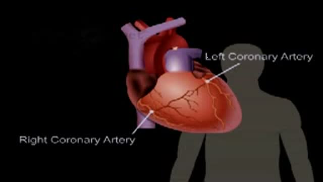
This patient-education video explains the anatomy and function of a healthy heart. The narration describes the four chambers, the heart's own coronary blood supply, and how it pumps blood around the body. It was produced as an introduction shown before a heart procedure. The video runs about a minute and a half in total.
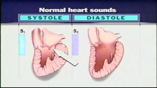
Normal Heart Sounds With the aid of a stethoscope you can hear the characteristic sounds of the normal heartbeat, typically described as a "lub-dub." These sounds are produced by the closure of the heart valves. The first heart sound or "lub" results from closure of the tricuspid and mitral valves. It is a rather low-pitched and a relatively long sound which, as indicated in, represents the beginning of ventricular systole. The second heart sound, or "dub," marks the beginning of ventricular diastole. It is produced by closure of the aortic and pulmonary (pulmonic) semilunar vanes when the intraventricular pressure begins to fall. This "dub" sound is typically heard as a sharp snap because the semilunar valves tend to close much more rapidly than the AV valves. Because diastole occupies more time than systole, a brief pause occurs after the second heart sound when the heart is beating at a normal rate. Therefore, the pattern that one hears is one of: "lub-dub" pause, "lub-dub" pause, and so on. Sometimes, especially in young normal individuals, a third heart sound can be heard. This sound is produced by the very rapid influx of blood into the partially filled ventricle. It is typically very faint and as such difficult to hear.
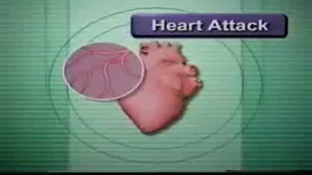
How to deal with heart attack and with stroke
Examination of the heart from the USMLE collection
This cardiology teaching clip explains how to hear the S3 and S4 heart sounds. The examiner shows how rolling the patient partly onto the left side and using the stethoscope bell with light pressure brings out low-pitched sounds. It supports training in cardiac auscultation.
This auscultation teaching clip guides the listener through the heart sounds S1 and S2 at each chest listening area. It explains where S2 is normally louder, the inspiratory splitting of S2 into its aortic and pulmonic components, and why S1 is usually the louder sound at the mitral area. It supports training in cardiac auscultation.
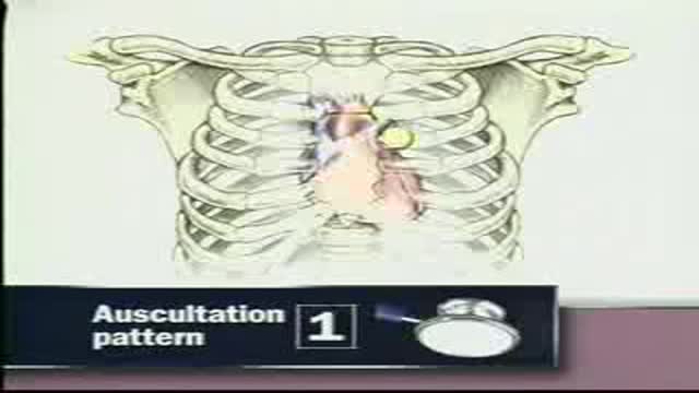
Auscultation of the heart is demonstrated in this cardiology examination video. The recording shows where the stethoscope is placed to listen to heart sounds during a cardiac examination. It is an educational resource for medical students learning the fundamentals of cardiac physical assessment.
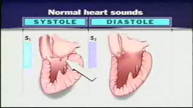
Heart sounds are reviewed in this cardiology listening exercise for clinical examination training. The recording works through the sounds heard with a stethoscope during assessment of the heart. It is a study resource for students learning cardiac auscultation basics and examination technique.
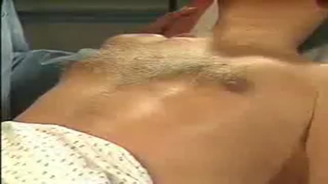
Examination of the heart is demonstrated in this cardiology video for clinical assessment training. The presenter works through the steps used to assess the heart during a physical examination. It is a study resource for students learning cardiac examination skills. The material is intended for general medical education and classroom study.
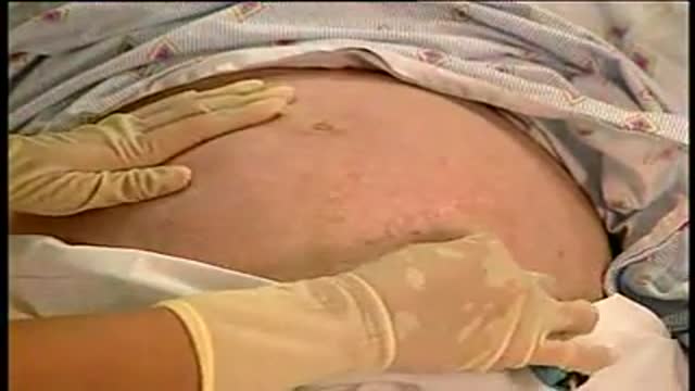
Fetal heart sounds recorded with a handheld monitor are presented in this short cardiology clip. Listening to the fetal heartbeat is a routine part of prenatal assessment, used to check the baby's rate and rhythm before birth. A brief learning clip for students. For learning purposes only — not medical advice.
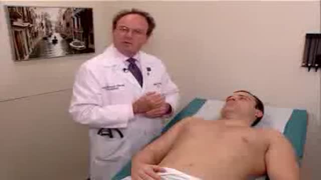
Examination of the heart and lungs is demonstrated in this clinical skills video, including listening to heart sounds. The presenter works through the standard steps used to assess the chest during a physical examination. The recording is a training resource for students learning cardiopulmonary examination technique.

This cardiology video demonstrates the heart examination in a child, covering the key steps of pediatric cardiac assessment. The examination includes inspection and palpation of the chest and listening to the heart sounds with a stethoscope. Heart examination is a routine part of child health assessments.
Medical Videos
Free medical and surgery videos for patients, students and professionals — trusted since 2007.
Founded by Dr. Mohamed Ibrahim, MD, PhD — physician-scientist.
Follow us on Facebook · Instagram