- Physical Examination
- Surgical Examination
- Ophthalmology
- Clinical Skills
- Orthopedics
- Surgery Videos
- Laparoscopy
- Pediatrics
- Funny Videos
- Cardiothoracic Surgery
- Nursing Videos
- Plastic Surgery
- Otorhinolaryngology
- Histology and Histopathology
- Neurosurgery
- Dermatology
- Pediatric Surgery
- Urology
- Dentistry
- Oncology and Cancers
- Anatomy Videos
- Health and Fitness
- Radiology
- Anaesthesia
- Physical Therapy
- Pharmacology
- Interventional Radiology
- Cardiology
- Endocrinology
- Gynecology
- Emergency Medicine
- Psychiatry and Psychology
- Childbirth Videos
- General Medical Videos
- Nephrology
- Physiology
- Diet and Food Health
- Diabetes Mellitus
- Neurology
- Women Health
- Osteoporosis
- Gastroenterology
- Pulmonology
- Hematology
- Rheumatology
- Toxicology
- Nuclear Medicine
- Infectious Diseases
- Vascular Disease
- Reproductive Health
- Burns and Wound Healing
- Kidney Zone Podcast
- Other
Carotid Arteries
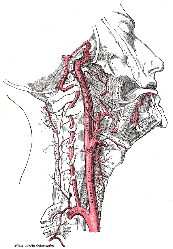
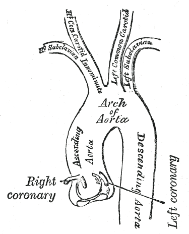
Gray's Anatomy (1918) plate, public domain
The two common carotid arteries ascend on either side of the windpipe, each dividing at the level of the jaw angle into the external carotid (supplying face, scalp and mouth) and the internal carotid, which enters the skull to supply most of the brain and the eye. At the division sits the carotid sinus, a pressure sensor that helps control blood pressure, and the carotid body, a chemical sensor for oxygen. The carotid pulse is palpable beside the windpipe.
Key facts
- The internal carotid has no branches in the neck — all neck branches come from the external carotid.
- The two internal carotids, with the vertebral arteries, form an arterial ring (circle of Willis) at the brain's base.
- Pressing the carotid sinus can slow the heart — the basis of carotid massage sometimes used in rhythm assessment.
- Turbulent flow through a narrowed carotid can be heard with a stethoscope as a bruit.
What can go wrong
Narrowing of the internal carotid by atherosclerosis at its origin is a major cause of stroke and transient ischaemic attacks (TIAs), including brief visual loss in the eye on the same side. Assessment uses ultrasound (carotid Doppler) and brain imaging; significant symptomatic narrowing is sometimes treated with surgery (endarterectomy) or stenting alongside medication. The carotid can also be injured or torn (dissection) after neck trauma or spontaneously, causing stroke in younger people.
Related conditions
Related procedures & tests
Related symptoms
Related muscles
Frequently asked questions
What is a carotid bruit?
A whooshing sound heard over the neck from turbulent blood flow, often through a narrowed artery; it prompts ultrasound assessment but can also occur without significant narrowing.
Why do carotid problems affect the eye?
The ophthalmic artery is the internal carotid's first branch inside the skull, so debris from carotid plaque can lodge in the retinal circulation, causing temporary or permanent visual loss.
Educational information only. This atlas explains normal anatomy and describes conditions in general terms; it is not medical advice and does not replace assessment by a qualified clinician. If you have symptoms that worry you, seek medical care.