- Physical Examination
- Surgical Examination
- Ophthalmology
- Clinical Skills
- Orthopedics
- Surgery Videos
- Laparoscopy
- Pediatrics
- Funny Videos
- Cardiothoracic Surgery
- Nursing Videos
- Plastic Surgery
- Otorhinolaryngology
- Histology and Histopathology
- Neurosurgery
- Dermatology
- Pediatric Surgery
- Urology
- Dentistry
- Oncology and Cancers
- Anatomy Videos
- Health and Fitness
- Radiology
- Anaesthesia
- Physical Therapy
- Pharmacology
- Interventional Radiology
- Cardiology
- Endocrinology
- Gynecology
- Emergency Medicine
- Psychiatry and Psychology
- Childbirth Videos
- General Medical Videos
- Nephrology
- Physiology
- Diet and Food Health
- Diabetes Mellitus
- Neurology
- Women Health
- Osteoporosis
- Gastroenterology
- Pulmonology
- Hematology
- Rheumatology
- Toxicology
- Nuclear Medicine
- Infectious Diseases
- Vascular Disease
- Reproductive Health
- Burns and Wound Healing
- Kidney Zone Podcast
- Other
Ear
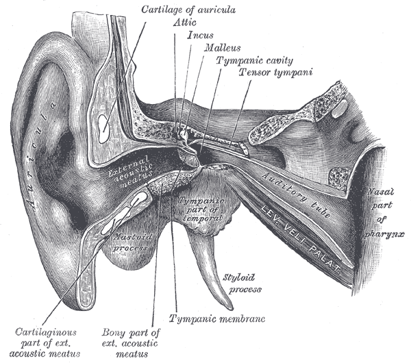
Gray's Anatomy (1918) plate, public domain
The ear has three parts. The outer ear collects sound and funnels it down the canal to the eardrum. The middle ear — an air-filled cavity in the temporal bone — holds the three ossicles that amplify vibrations. The inner ear contains the cochlea, which converts vibration into nerve signals, and the balance organs (vestibular system) that sense head movement.
Key facts
- The ear canal is about 2.5 centimetres long and slightly S-shaped.
- The middle ear is ventilated by the Eustachian tube from the back of the nose.
- The cochlea is a spiral tube about 35 millimetres long, wound like a snail shell.
- The balance organs are three semicircular canals set at right angles to each other.
- Hearing and balance share the vestibulocochlear nerve.
What can go wrong
Outer-ear problems include wax blockage and infection of the canal; middle-ear problems include infection (especially in children), fluid behind the drum (glue ear) and ossicle problems. Inner-ear conditions cause hearing loss, tinnitus and vertigo. Ear pain can also be referred from the jaw joint, teeth or throat.
Related procedures & tests
- Audiometry (hearing test)
- Ear wax removal (microsuction / irrigation)
- Ear grommet insertion (myringotomy with tubes)
Related symptoms
Watch: related videos
Frequently asked questions
Why do ears pop on aeroplanes?
Pressure changes faster outside than in the middle ear; the Eustachian tube must open to equalise it. Swallowing or yawning opens the tube — the pop is the pressure balancing.
Is earwax bad?
No — wax protects and cleans the canal, migrating outwards on its own. Problems come from blockage, often worsened by cotton buds pushing wax deeper.
Educational information only. This atlas explains normal anatomy and describes conditions in general terms; it is not medical advice and does not replace assessment by a qualified clinician. If you have symptoms that worry you, seek medical care.