- Physical Examination
- Surgical Examination
- Ophthalmology
- Clinical Skills
- Orthopedics
- Surgery Videos
- Laparoscopy
- Pediatrics
- Funny Videos
- Cardiothoracic Surgery
- Nursing Videos
- Plastic Surgery
- Otorhinolaryngology
- Histology and Histopathology
- Neurosurgery
- Dermatology
- Pediatric Surgery
- Urology
- Dentistry
- Oncology and Cancers
- Anatomy Videos
- Health and Fitness
- Radiology
- Anaesthesia
- Physical Therapy
- Pharmacology
- Interventional Radiology
- Cardiology
- Endocrinology
- Gynecology
- Emergency Medicine
- Psychiatry and Psychology
- Childbirth Videos
- General Medical Videos
- Nephrology
- Physiology
- Diet and Food Health
- Diabetes Mellitus
- Neurology
- Women Health
- Osteoporosis
- Gastroenterology
- Pulmonology
- Hematology
- Rheumatology
- Toxicology
- Nuclear Medicine
- Infectious Diseases
- Vascular Disease
- Reproductive Health
- Burns and Wound Healing
- Kidney Zone Podcast
- Other
Lumbar Vertebrae
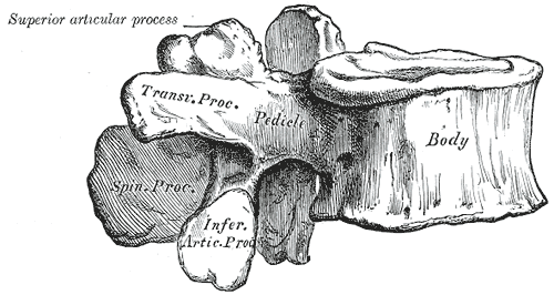
Gray's Anatomy (1918) plate, public domain
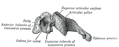
The five lumbar vertebrae are the largest in the spine, forming the small of the back. Their massive bodies bear the weight of the head, arms and trunk, and their chunky processes anchor the powerful back and abdominal-wall muscles. Between them sit the thickest intervertebral discs, and below them the spinal cord has tapered into a bundle of nerves.
Key facts
- There are five lumbar vertebrae, L1 to L5.
- They form the forward curve (lordosis) of the lower back.
- Their bodies are the largest of any vertebrae, for load-bearing.
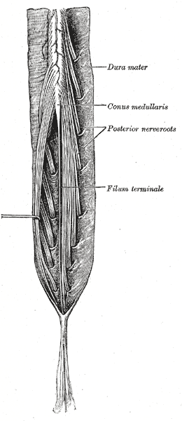
- The spinal cord ends around L1-L2; below that the canal holds loose nerve roots (the cauda equina).
- Lumbar punctures are done below L2, between these vertebrae, to avoid the cord.
What can go wrong
The lumbar region is the commonest site of back pain: discs here can bulge or prolapse and press on nerve roots, causing sciatica. Wear in the facet joints and narrowing of the canal (spinal stenosis) can make walking distances shrink with age. Fractures are usually from significant trauma or osteoporosis.
Related procedures & tests
Related symptoms
Related muscles
Watch: related videos
Frequently asked questions
Why are lumbar vertebrae so big?
Load. Each one carries the weight of everything above it plus the forces of lifting, so their bodies are wide, kidney-shaped blocks of bone — the largest in the column.
What is the cauda equina?
Latin for 'horse's tail': below the end of the spinal cord, the canal contains a loose bundle of nerve roots that resemble a tail. New bladder or bowel problems with back pain and saddle numbness can signal pressure on these nerves and need emergency assessment.
Educational information only. This atlas explains normal anatomy and describes conditions in general terms; it is not medical advice and does not replace assessment by a qualified clinician. If you have symptoms that worry you, seek medical care.