
- Physical Examination
- Surgical Examination
- Ophthalmology
- Clinical Skills
- Orthopedics
- Surgery Videos
- Laparoscopy
- Pediatrics
- Funny Videos
- Cardiothoracic Surgery
- Nursing Videos
- Plastic Surgery
- Otorhinolaryngology
- Histology and Histopathology
- Neurosurgery
- Dermatology
- Pediatric Surgery
- Urology
- Dentistry
- Oncology and Cancers
- Anatomy Videos
- Health and Fitness
- Radiology
- Anaesthesia
- Physical Therapy
- Pharmacology
- Interventional Radiology
- Cardiology
- Endocrinology
- Gynecology
- Emergency Medicine
- Psychiatry and Psychology
- Childbirth Videos
- General Medical Videos
- Nephrology
- Physiology
- Diet and Food Health
- Diabetes Mellitus
- Neurology
- Women Health
- Osteoporosis
- Gastroenterology
- Pulmonology
- Hematology
- Rheumatology
- Toxicology
- Nuclear Medicine
- Infectious Diseases
- Vascular Disease
- Reproductive Health
- Burns and Wound Healing
- Kidney Zone Podcast
- Other
Small bowel MRI (MR enterography)
Category: Digestive Tests & Procedures · All digestive tests & procedures
What it is
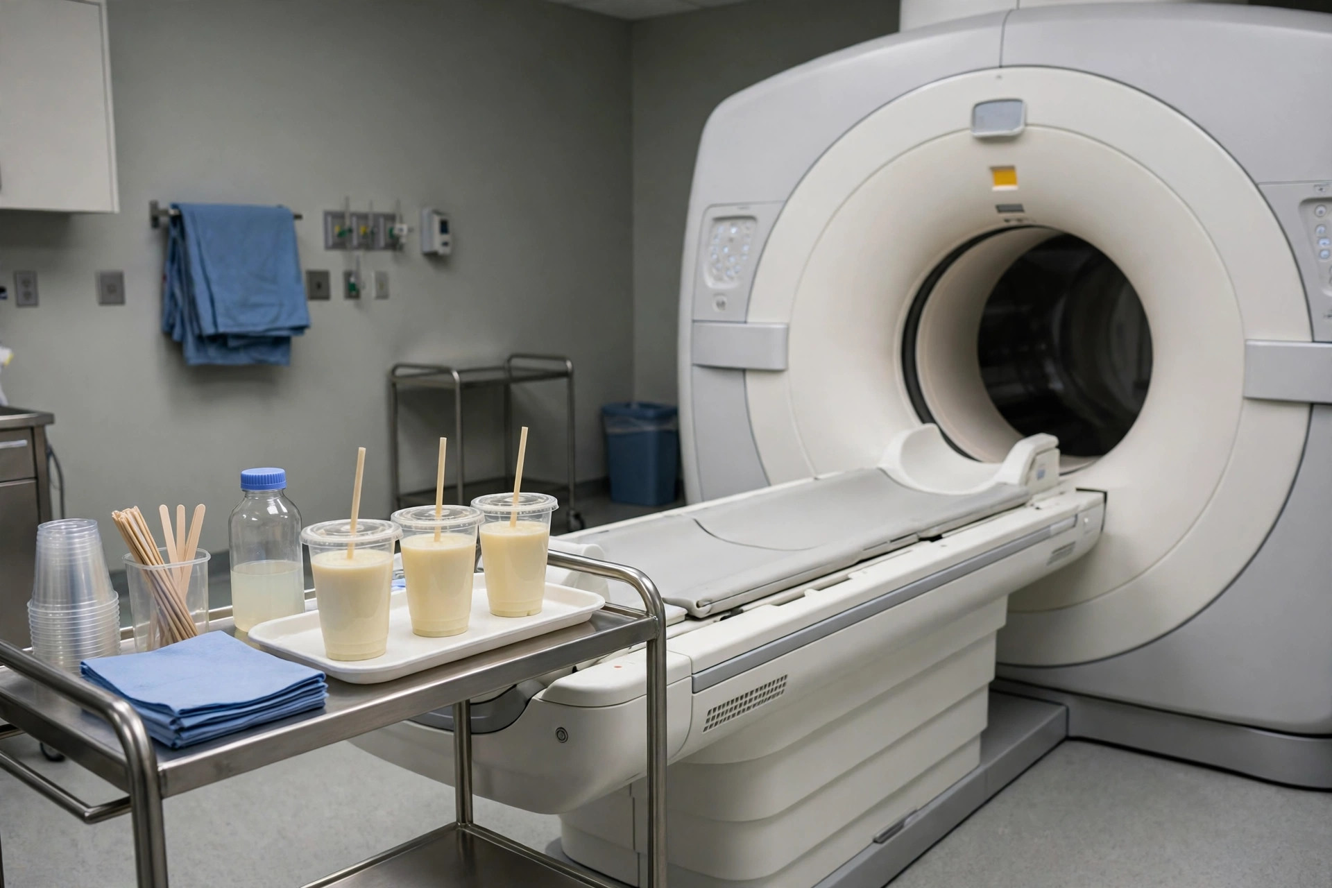
MR enterography is an MRI scan focused on the small bowel. You drink a special fluid that distends the bowel so its wall can be assessed for inflammation, narrowing and other abnormalities, with no radiation involved.
Why it is done
It is mainly used in Crohn's disease — for diagnosis, mapping its extent and checking treatment response — and for other small-bowel problems when repeated imaging without radiation is valuable.
How to prepare
You will fast for several hours and then drink a measured volume of the distending fluid over about 45 minutes before the scan; it can cause loose stools later. Standard MRI metal-safety checks apply. Tell the team about kidney problems and allergies.
What happens during the procedure
During the scan (about 30 to 45 minutes) you lie in the MRI scanner, hold your breath for short sequences, and may be given an injection to relax the bowel and a contrast dye through a vein. The scanner is noisy and you wear ear protection.
Afterwards — recovery & results
You can go home afterwards. The drinks commonly cause diarrhoea for some hours, which is expected. The report is sent to your specialist team, usually within days.
Risks & considerations
There is no ionising radiation. Risks include discomfort from the bowel drinks, a small chance of reaction to the contrast dye, and the usual MRI cautions about implants and metal.
Related guides on MedicalVideos.com
Frequently asked questions
Why drink so much fluid?
A collapsed bowel hides its wall. The drink inflates the small bowel so thickened or inflamed sections stand out clearly.
Why MRI rather than CT for Crohn's?
People with Crohn's often need repeated imaging over many years. MRI gives similar detail without radiation, so it is preferred for monitoring.
Related procedures — Digestive Tests & Procedures
Browse the S section of the A–Z list or return to the Procedures & Tests hub.