
- Procedures & Tests
- Symptoms A-Z
- Symptom Checker
- Kidney Diet
- First Aid
- Anatomy Atlas
- Ask MedicalVideos
- Drug Guide
- Lab Values
- Vital Signs
- Muscles A–Z
- Clinical Signs
- Medical Abbreviations
- Medical Terminology
- Physical Examination
- Surgical Examination
- Ophthalmology
- Clinical Skills
- Orthopedics
- Surgery Videos
- Laparoscopy
- Pediatrics
- Funny Videos
- Cardiothoracic Surgery
- Nursing Videos
- Plastic Surgery
- Otorhinolaryngology
- Histology and Histopathology
- Neurosurgery
- Dermatology
- Pediatric Surgery
- Urology
- Dentistry
- Oncology and Cancers
- Anatomy Videos
- Health and Fitness
- Radiology
- Anaesthesia
- Physical Therapy
- Pharmacology
- Interventional Radiology
- Cardiology
- Endocrinology
- Gynecology
- Emergency Medicine
- Psychiatry and Psychology
- Childbirth Videos
- General Medical Videos
- Nephrology
- Physiology
- Diet and Food Health
- Diabetes Mellitus
- Neurology
- Women Health
- Osteoporosis
- Gastroenterology
- Pulmonology
- Hematology
- Rheumatology
- Toxicology
- Nuclear Medicine
- Infectious Diseases
- Vascular Disease
- Reproductive Health
- Burns and Wound Healing
- Kidney Zone Podcast
- Other

Thyroid Swelling

Educational only: videos on MedicalVideos.com are for general information and are not medical advice. Always consult your own doctor about your health.
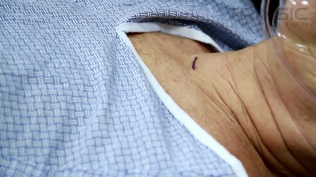
Hypothyroidism and Hyperthyroidism Symptoms: Swollen Neck. A goiter is an enlargement of the thyroid gland. As shown here, an enlarged thyroid can be seen as a swelling in the front of the neck. A goiter can occur with either hypothyroidism or hyperthyroidism
This video in 30 seconds
A classic surgical-teaching lecture demonstrates examination of a thyroid swelling. It covers inspection with the neck extended and the patient swallowing, palpation and measurement of the swelling against nearby landmarks, and percussion and auscultation. The wider examination follows, including eye signs, tremor, pulse and signs related to thyroid function and possible spread.
Key points
- Inspection includes watching the swelling move on swallowing and tongue movement
- Palpation defines the lobes, borders, consistency and any nodules
- Percussion and auscultation check for retrosternal extension and bruits
- Eye signs, tremor and pulse are examined for thyroid overactivity
- Signs of underactivity and of metastasis are reviewed at the end
Chapters
- 0:02 — Inspecting the thyroid swelling
- 5:25 — Palpating the thyroid gland
- 10:41 — Trachea and carotid assessment
- 14:24 — Percussion and auscultation
- 15:41 — Eye signs of thyrotoxicosis
- 21:02 — Tremor, pulse and general signs
- 26:11 — Checking for metastasis
Video Transcript
A classic surgical-teaching lecture on examining a thyroid swelling, from the same series as the swelling-examination video. It covers inspection with the neck extended and the patient swallowing, measuring the swelling and defining its borders against the sternomastoid and the suprasternal notch, palpation, and the wider examination — lymph nodes, the skull and long bones, the abdomen and the chest — before the case is concluded.
Read the full transcript
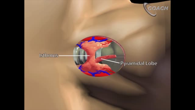
Examination of thyroid swelling The thyroid gland is located in front of the neck with its two lobes on either sides of the trachea connected by anysthmus. We begin our examination of the thyroid swelling by inspection. For inspection, make the patient sit on a stool with the neck slightly hyperextended and inspect the thyroid from the front. Asking the patient to swallow makes the thyroid more prominent for inspection. If the patient has a short and fat neck, ask the patient to clasp the hands over the occipit and push the head backwards against the resistance of the clasped hands.
This is the pisillos method to make the thyroid more prominent for inspection. First, note the size and shape of the swelling and its situation, whether it is on one side of the midline, in the midline or whether it extends on both the sides. Measure the exact size in centimeters using a measuring tape. Now note the extent of the swelling, particularly the lateral border in relation to the sternomastoid and the lower border in relation to the suprasternal notch of the sternum. A thyroid swelling always lies deep to the sternomastoid muscles as demonstrated here by contracting the sternomastoids.
Then note whether the lower border ends above the suprasternal notch or it extends behind the suprasternal notch. Both the borders will be better appreciated if the patient is asked to make swallowing movements during the inspection. Next, inspect the surface of the swelling. A simple goiter and single nodules have a smooth surface. While a multinaudial goiter has a nodular or bossillated surface. Note the bossillated surface of this large multinaudial goiter in a young male patient. Then inspect the skin over the swelling and look for redness and edema, suggestive of inflammation, scars of previous surgery, sinuses and dilated veins.
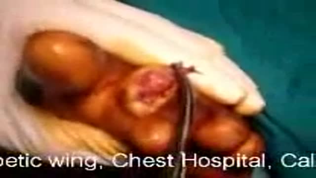
Note this transverse scar of previous thyroid surgery which tells us that this is a recurrent thyroid swelling. And this is a sinus in the midline of the neck discharging mucus like fluid. This is a thyroglossal fistula. Then watch the swelling for a few seconds quietly for pulsatility. Now ask the patient to swallow and look for an upward movement of the swelling. A thyroid swelling characteristically acts during the glutation as it is enclosed in the pre tracheal fascia which is fixed to the thyroid cartilage. So when the superior constrictors of larynx pull the thyroid cartilage up during the glutation, the thyroid also moves up and down.
Of course we should not forget the other swellings that also move on the glutation. That is the thyroglossal fists, pre tracheal lymph nodes, sub hyoid bursa and extrinsic carcinoma of larynx. A swelling which is not attached to the pre tracheal fascia like this lipoma will not move upwards during the glutation. Note the upward movement of the thyroid cartilage but not of the swelling. If the swelling is a nodule close to the midline, you must also test for its upward movement on protrusion of the tongue. Ask the patient to extend the neck and open the mouth wide.
Now keeping the mouth open, let the patient move the tongue out and in. This is a thyroglossal cyst and note the upward movement of the swelling as the tongue is protruded. This sign is diagnostic of a thyroglossal cyst which is connected to the foramen cecum of the tongue. A thyroglossal fistula also moves upwards on protrusion of the tongue as the fistula is connected to the foramen cecum. And of course it also moves up with the glutation. To revise on inspection, first note the size and shape of the swelling, its location and its borders in relation to the sternomastoid and the suprasternal notch.
Then inspect the surface whether smooth, nodular or bossillated. Then inspect the skin over the swelling for redness and edema, scars and sinuses and dilated veins. Look for any visible pulsations in the swelling. Then ask the patient to swallow and look for an upward movement of the swelling during the glutation. And lastly if the swelling is close to the midline, look for an upward movement of the swelling on protruding the tongue. Now let us proceed to palpation. After completing inspection, let the patient be in the same position sitting with neck extended and test the temperature of the swelling with the back of the fingers and then look for tenderness.
Then stand behind the patient, place the hands around the neck with the thumbs over the occiput and tips of the other fingers over the front of the neck. This is the standard method for palpation of the thyroid gland. The flexion of the neck can be adjusted by the grip between the thumbs over the occiput and the index fingers under the chin. Now keep the neck slightly flexed to relax the deep cervical fascia and ask the patient to make a swallowing movement as you are palpating. As the thyroid moves up and down, the lower border can be appreciated better and nodules if present can be felt more easily.
Then palpate the anterior surfaces of the lobes one at a time, inclining the head to the side being examined to relax the over-links sternum asteroid muscle. For example, to examine the right lobe, flex the neck slightly to the right, support the thyroid firmly with the left hand and then palpate with the right hand. Note the consistency, surface and borders carefully. Palpate the tracheal rings in the suprastermal notch, that is below the lower border of the isthmus. If they are not felt, a retrosterinal extension should be suspected.
Now stand in front of the patient, extend the neck slightly and palpate by Lahis method for deep or posterior medial surface of the gland. To palpate the left lobe, push the thyroid gland to the left by your left hand so that the left lobe is pushed out of the tracheoisophageal groove and then with the right hand palpate the posterior surface for nodules. Then repeat the procedure on the right side. Next, with the patient in the same position, keep a thumb over the lobe to be examined and ask the patient to swallow.
Feel for small nodules on the surface as the gland moves up and down. This is Kryl's method of palpation for small nodules. During palpation, first you should determine whether the entire thyroid gland is enlarged or is it an affection of only one lobe or is it a single nodule. If an entire gland or an entire lobe is palpable, note its surface whether it is smooth or bossillated and its consistency whether it is uniform or variable, soft, firm or hard. Colloid goiters are soft, multinodular goiters are firm and carcinoma and riddles thyroiditis are hard to feel.
Secondly, palpate the lower border very carefully during deglutition to determine the presence of a retrosternal extension. Palpation of tracheal rings above the suprasternal notch rules out a retrosternal extension. If it is a single nodule, note its position in the thyroid lobe or isthmus. Note its shape, measure its size and feel the consistency. A paradox about the consistency of thyroid nodule should be noted and remembered here that a cyst in thyroid feels firm while a solid swelling that is adenoma feels soft. Because adenomas being highly cellular are soft, while cysts with fluid under tension feel firm as compared to the normally soft thyroid.
After palpating the nodule, try to palpate the rest of the thyroid gland. Normally it is not palpable. A normal thyroid gland is not felt on palpation, only its isthmus can be felt over the second, third and fourth tracheal rings. So if along with a single nodule, like this nodule on the isthmus, the rest of the thyroid gland is palpable, clinically it is considered as a multi-nodular goiter with a single large nodule. But if the rest of the gland is not palpable, as in this case, then it is considered as a solitary nodule.
Now keep your hand very gently over the upper pole of each side and try to feel for a palpable thrill. Thrill over the upper pole akin to the feel of purring of a cat is diagnostic of a primary toxic goiter. Next, pinch the skin over the swelling to look for fixity to skin and test the mobility of the swelling in vertical and horizontal directions. Fixity in any direction suggests malignant infiltration or thyroiditis. Now, pulpate for the trachea and carotid pulsations. To pulpate the trachea, first note the position of the larynx and pulpate the trachea downwards.
In this unilateral left-sided goiter, the trachea can be felt along its length and its displacement to the opposite side can be easily appreciated. In a bilateral goiter, the trachea may be difficult to feel. So note the position of the larynx and the position of the trachea over the suprasternal notch to get an approximate idea of the position of the trachea. Then perform the Cocker's Test to rule out tracheal narrowing. Ask the patient to extend the neck and take heavy deep breaths to open mouth. Now, compress the swelling from both the sides.
Appearance of strider on slight compression of the lateral lobes indicates narrowing of the trachea and this is a positive Cocker's Test. Listen to the strider on compression of the lateral lobes. This is typically seen in the scabar trachea of large and long-standing multinodular goiters, where trachea is compressed from both the sides and becomes an anterior posterior slit like the scabar of a sword. So lateral compression immediately produces obstruction and strider. It may also be seen in a malignant thyroid infiltrating the trachea. Now, pulpit the carotid artery pulsations against the transverse process of the sixth cervical vertebra between the posterior border of thyroid and the sternostoid.
In a unilateral goiter, first pulpit the normal side for normal feel of the carotid pulsation and then compare it with the affected side. If the goiter is large, the carotid sheath will be pushed backwards, but the pulsations will always be well-felt in spite of the backward displacement in a benign goiter. But in a malignant goiter, the carotid pulsations may be felt weak or obliterated due to malignant infiltration of the carotid sheath. This is a positive, various sign, that is obliteration of carotid pulsation in malignant thyroid. To revise, we pulpit by the standard method, Lahe's method and Kreil's method.
Note on pulpation the exact size and shape of the swelling and identify its borders. In particular, pulpit the tracheal rings in the suprasternal notch to identify a retrosternal extension, if any. Then note the surface and the consistency of the swelling and pulpit carefully by Lahe's method and Kreil's method for small nodules within the gland. In case of a single nodule, note whether the rest of the gland is palpable. Now look for a palpable thrill over the upper pole and for fixity to skin or deeper structures. Lastly, pulpit the trachea and the carotids.
Trace the trachea, look for deviation from the midline and if goiter is large or bilateral, perform the cocker's test. Lastly, pulpit the carotids behind the posterior border of the swelling. Now let us proceed to percussion and auscultation. Percuss directly over the manubrium sternum or use a heavy percussion stroke. Normally the note is resonant due to air in the trachea behind the manubrium sternum. If the percussion note is dull, suspect a retrosternal extension of thyroid which descends between the trachea and the sternum and comes to light between the manubrium sternum and the air column.
Auscultation. Auscultate over the swelling paying more attention at the upper poles for a systolic bruit. Bruit is diagnostic of primary toxic goiter and is due to markedly increased vascularity of the gland. Having completed the examination of the swelling and concluded that it is a thyroid swelling, now you should look specifically for signs of thyrotoxicosis, signs of myxidema, signs of retrosternal extension and signs of metastasis. Signs of thyroidoxicosis. First, let us look for the eye signs. Ask the patient sitting upright on a stool to look horizontally forwards towards a distant object at eye level.
Normally the lower lid just touches the limbus at 6 o'clock while the upper lid covers the upper margin of the cornea from 11 to 1 o'clock. Limbus is the corneal margin. The early eye signs of thyrotoxicosis are lid retraction, lid lag and infrequent blinking. The earliest eye sign to appear is the lid retraction with resultant widening of the palpabral fissure, also termed as daldrympel sign. Note here the lid retraction which is more prominent in the left eye. The upper lid is retracted due to tonic contraction of the involuntary part of the levator palpabrae superioris,
thereby exposing a strip of white sclera above the superior limbus. This results in a typical staring and frightened look of a thyrotoxic patient. This is associated with infrequent and incomplete blinking of the eyes, termed as stalwax sign. Note how infrequently the patient blinks and when she blinks it is only partial. Now test for lid lag or one graphe sign. With the left hand fix the patient's head to prevent neck flexion or extension. Hold your right index finger in front of the patient's eye at a distance of 40 cm
and asking the patient to follow the finger move it upwards and downwards. When the patient looks downwards the upper lid lags behind and a larger portion of the white sclera is exposed on looking down. This is a positive one graphe sign. With further progress of thyrotoxicosis there is actual bulging of the eyeballs due to retroorbital edema and fat deposition. This is termed as exopthelmos. Ask the patient to stare horizontally forwards and a strip of white sclera is visible between the inferior limbus and the lower eyelid. This has nothing to do with the upper lid retraction and it is due to actual forward bulging of the eyeball that is exopthelmos.
Now to note the amount or degree of exopthelmos examine the eyes by Nefziger's method. Stand behind the patient. Tilt the head of the patient backwards to observe the eyes in supra orbital plane. The actual protrusion of the eyeballs beyond the supra orbital ridges can be appreciated in this position. Normally the eyes do not project beyond the supra orbital ridges. Now ask the patient to look upwards with head tilted slightly downwards. Look for the transverse wrinkles over the forehead which are seen in a normal individual in this position.
Absence of wrinkling of forehead in thyrotoxicosis is positive joffroys sign. If the exopthelmos is limited to one eye it has to be differentiated from proptosis due to an intra orbital mass. Ask the patient to look downwards and even the upper eyelid. The aversion is difficult or impossible in exopthelmos due to thyrotoxicosis while in a case of intra orbital mass aversion is possible even with very marked proptosis. This is the Gifford sign. Now test convergence by holding your finger at a distance of one meter from the eyes and asking the patient to stare at it.
Now bring it slowly towards the midpoint between the eyebrows. The patient cannot converge the eyes in exopthelmos. This is a positive mobius sign. Convergence is slow, jerky and incomplete. With progressive exopthelmos there is further bulging of the eyeballs with conjunctival congestion and edema. In a later stage there are corneal ulcers, diminished vision and ophthalmoplegia or restriction of the eyeball movements. To revise look for lead retraction, lead lag and infrequent blinking. Look for actual exopthelmos by Nefziger's method and visibility of sclera beneath the inferior limbus. Then look for wrinkling of forehead that is Joffroy's sign and test convergence that is Mobius sign.
We are examining for the signs of thyrotoxicosis and we have now studied the eye signs in thyrotoxicosis. Next look for fine tremors of outstretched hands and protruded tongue. Ask the patient to stretch out the arms with slight fanning of the fingers. Thyrotoxic patients will always show a fine tremor of the fingers. This is another patient with a thyroid enlargement. Note the fine tremor of the fingers of the outstretched hands suggesting thyrotoxicosis. Another method to demonstrate tremors is to keep a piece of paper over the outstretched fingers.
The vibrations of the paper are more easily appreciated. Then ask the patient to open the mouth fully and put out the tongue straight for 15 to 30 seconds. The tongue should not touch the teeth. Look for frank tremors or in mild cases fine febrile-ray twitchings. Note again the tremors of the protruded tongue in thyrotoxicosis. Next feel the radial pulse. Tachycardia and bounding pulse are characteristic of thyrotoxicosis. However, pulse rate may increase further due to anxiety which is associated with thyrotoxicosis. So to judge the degree of thyrotoxicosis you must count the sleeping pulse rate
which is counted early morning around 4 a.m. that is before the patient wakes up without disturbing the sleep. A fast and irregular pulse may suggest atrial fibrillation. Now feel the pulse and feet of the patient. In thyrotoxicosis they are warm and moist and the legs may show pre-tibial mixidema with thickened hyperpigmented skin and coarse hair. Lastly another characteristic sign of thyrotoxicosis has already been studied by us during the examination of the goiter that is the palpable thyroid thrill and thyroid gruy on oscultation. Let us remember here that in primary thyrotoxicosis the eye signs and tremors are more prominent
while in secondary thyrotoxicosis cardiac signs are more prominent with tachycardia, cardiomegaly, atrial fibrillation or congestive cardiac failure. In secondary thyrotoxicosis the tremors are less prominent and eye signs and exopthelmos are not seen. Signs of mixidema. If there are no signs of thyrotoxicosis look for signs of mixidema. Is the voice hoarse? Is there edema of the face and legs? Does the patient feel lethargic? And look for the diagnostic clinical sign that is delayed relaxation of the deep reflexes. Test the ankle jerk and the knee jerk. Note the typical mixidema phases with edema and dry coarse skin and listen to the characteristic slow hoarse voice.
Now note the slow relaxation phase of this ankle jerk. The muscle contracts briskly as usual and then relaxes slowly. In this early case note the slightly delayed relaxation phase of the ankle jerk. Now we look for the signs of retrosternal extension. Inspect the upper chest, neck and face for dilated veins, congestion and puffiness which result from pressure on the internal jugular veins at the thoracic inlet. Pulpate the tracheal rings in the suprasternal notch. Inability to pulpate the rings suggests a retrosternal goiter. Then percuss over the manubrium sterni.
A dull note instead of normal resonant note suggests the presence of a retrosternal goiter. Now elevate both arms above the head until they touch the sides of the face. Hold them there for one minute. If this manure produces congestion of the face and respiratory distress, it suggests thoracic outlet obstruction which in case of a goiter is most likely to be due to retrosternal extension of the goiter. This is positive pamperton sign. Lastly look for Horner's syndrome which can occur in a retrosternal goiter or in a malignant goiter affecting the cervical sympathetic nerve.
The features of Horner's syndrome are ptosis with constricted pupil and inoc thalamus in the eye of the affected side. The ciliospinal reflex is lost and there is anhydrosis or loss of sweating on the affected side of the face. Lastly look for the evidence of metastasis meticulously if carcinoma is suspected. Pulpate the neck on both sides for enlarged hard lymph nodes. Pulpate the entire skull surface for hard nodules. Look for bony deformity or tenderness in long bones. Examine the abdomen for nodular liver and acitis. And examine the chest for signs of effusion and consolidation.
This concludes the clinical examination of a case of thyroid swelling.
Transcript auto-generated from the video’s audio for accessibility and study. Educational content only — not medical advice.
Related reading
Medical Videos
Free medical and surgery videos for patients, students and professionals — trusted since 2007.
Founded by Dr. Mohamed Ibrahim, MD, PhD — physician-scientist.
Follow us on Facebook · Instagram
SORT BY-
Top Comments
-
Latest comments