- Physical Examination
- Surgical Examination
- Ophthalmology
- Clinical Skills
- Orthopedics
- Surgery Videos
- Laparoscopy
- Pediatrics
- Funny Videos
- Cardiothoracic Surgery
- Nursing Videos
- Plastic Surgery
- Otorhinolaryngology
- Histology and Histopathology
- Neurosurgery
- Dermatology
- Pediatric Surgery
- Urology
- Dentistry
- Oncology and Cancers
- Anatomy Videos
- Health and Fitness
- Radiology
- Anaesthesia
- Physical Therapy
- Pharmacology
- Interventional Radiology
- Cardiology
- Endocrinology
- Gynecology
- Emergency Medicine
- Psychiatry and Psychology
- Childbirth Videos
- General Medical Videos
- Nephrology
- Physiology
- Diet and Food Health
- Diabetes Mellitus
- Neurology
- Women Health
- Osteoporosis
- Gastroenterology
- Pulmonology
- Hematology
- Rheumatology
- Toxicology
- Nuclear Medicine
- Infectious Diseases
- Vascular Disease
- Reproductive Health
- Burns and Wound Healing
- Kidney Zone Podcast
- Other
Fallopian Tubes
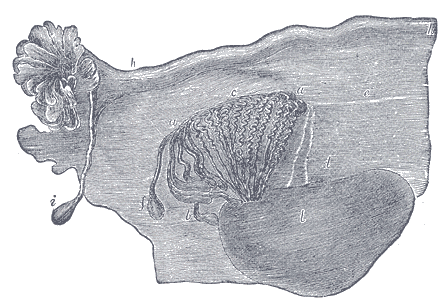
Gray's Anatomy (1918) plate, public domain
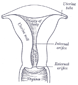
The fallopian tubes are two slender muscular tubes, about 10-12 cm long, arching from the top of the uterus towards the ovaries. Their fringed ends (fimbriae) sweep over the ovary to catch the released egg; tiny cilia and muscle waves then transport the egg towards the uterus. Fertilisation normally happens in the tube's outer third, and the early embryo travels down over several days.
Key facts
- The tube is not attached to the ovary — its fimbriae move to cup the ovary at ovulation.
- Cilia lining the tube beat in coordinated waves toward the uterus, assisted by gentle muscle contractions.
- The journey of a fertilised egg down the tube takes about 3-4 days, timed to arrive as it becomes ready to implant.
- Cells lining the tube secrete fluid that nourishes the egg and early embryo.
What can go wrong
Damage or blockage of the tubes — most often from pelvic infection (such as chlamydia), endometriosis or previous surgery — is a major cause of infertility. If a fertilised egg implants in the tube instead of the uterus, the result is an ectopic pregnancy, which cannot survive and can rupture the tube, causing dangerous internal bleeding. Tube patency is tested with dye studies such as the hysterosalpingogram (HSG).
Related conditions
Related procedures & tests
Related symptoms
Frequently asked questions
What is an ectopic pregnancy?
A pregnancy that implants outside the uterine cavity, usually in a fallopian tube. As it grows it can rupture the tube, so early pregnancy pain or bleeding needs prompt assessment.
How do doctors check whether the tubes are open?
By imaging tests that pass dye or contrast through the uterus and tubes (HSG or ultrasound-based tests), or directly at laparoscopy with dye.
Educational information only. This atlas explains normal anatomy and describes conditions in general terms; it is not medical advice and does not replace assessment by a qualified clinician. If you have symptoms that worry you, seek medical care.