- Physical Examination
- Surgical Examination
- Ophthalmology
- Clinical Skills
- Orthopedics
- Surgery Videos
- Laparoscopy
- Pediatrics
- Funny Videos
- Cardiothoracic Surgery
- Nursing Videos
- Plastic Surgery
- Otorhinolaryngology
- Histology and Histopathology
- Neurosurgery
- Dermatology
- Pediatric Surgery
- Urology
- Dentistry
- Oncology and Cancers
- Anatomy Videos
- Health and Fitness
- Radiology
- Anaesthesia
- Physical Therapy
- Pharmacology
- Interventional Radiology
- Cardiology
- Endocrinology
- Gynecology
- Emergency Medicine
- Psychiatry and Psychology
- Childbirth Videos
- General Medical Videos
- Nephrology
- Physiology
- Diet and Food Health
- Diabetes Mellitus
- Neurology
- Women Health
- Osteoporosis
- Gastroenterology
- Pulmonology
- Hematology
- Rheumatology
- Toxicology
- Nuclear Medicine
- Infectious Diseases
- Vascular Disease
- Reproductive Health
- Burns and Wound Healing
- Kidney Zone Podcast
- Other
Bile Ducts
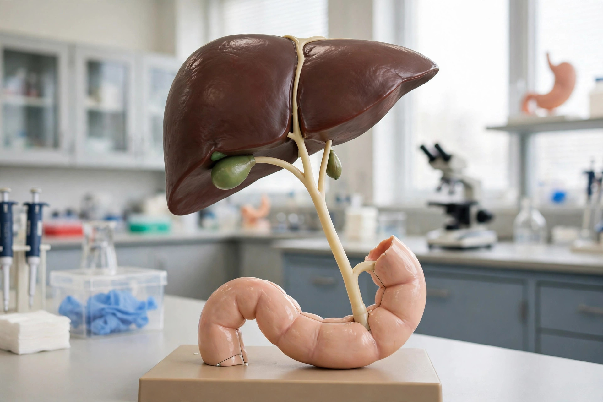
AI-generated anatomical illustration
The bile ducts are the drainage network carrying bile from the liver to the intestine. Small ducts within the liver merge into right and left hepatic ducts, join to form the common hepatic duct, receive the cystic duct from the gallbladder, and continue as the common bile duct, which empties into the duodenum alongside the pancreatic duct. Bile flow emulsifies dietary fat and carries waste products such as bilirubin out of the body.
Key facts
- The common bile duct is normally only about 6 mm wide — narrower than a pencil.
- Bile ducts are lined by cells that modify bile as it flows, adding bicarbonate and water.
- The sphincter of Oddi, a muscular valve where the ducts enter the duodenum, meters bile flow.
- The ducts' branching pattern varies; some people have accessory ducts that surgeons map carefully.
What can go wrong
Blockage is the central problem: a migrated gallstone, inflammation or a tumour can obstruct the duct, causing obstructive jaundice, pale stools, dark urine and itching; infection of a blocked duct (cholangitis) causes fever and rigors and is an emergency. Autoimmune diseases (primary biliary cholangitis, primary sclerosing cholangitis) scar the ducts over years. ERCP and MRCP imaging are used to examine and relieve blockages.
Related conditions
Related procedures & tests
Related symptoms
Frequently asked questions
Why does bile duct blockage cause itching?
When bile cannot drain, bile salts accumulate in the blood and deposit in the skin, where they are thought to trigger the intense itching of obstructive jaundice.
What is an MRCP?
A type of MRI scan that uses the fluid signal in the ducts to build a map of the bile and pancreatic ducts without any instruments entering the body.
Educational information only. This atlas explains normal anatomy and describes conditions in general terms; it is not medical advice and does not replace assessment by a qualified clinician. If you have symptoms that worry you, seek medical care.